Arslantunalı-Şahin vd. (2025), Mater Sci: Mater Med
Arslantunali-Sahin, D., Tamay, D.G., Isik, S. et al. (2025). Nerve tissue model on a micropatterned surface: Axon guidance and neural regeneration. J Mater Sci: Mater Med 37, 18.
DOI: https://doi.org/10.1007/s10856-025-06959-3
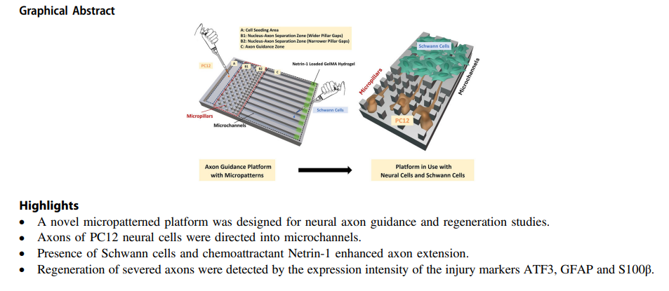
This study focuses on the design, production and testing of a micropatterned PDMS surface, featuring micropillars and microchannels to study the regeneration of individual axons of PC12 nerve cells after injury. Micropillar organization on the surface was designed to restrict the PC12 cell bodies while axons were guided into microchannels, allowing observation of individual axons. Surfaces were coated with poly(L-lysine) to improve cell attachment and proliferation. Netrin-1, a chemoattractant molecule and axonal elongation enhancer, was introduced in a gelatin methacrylate (GelMA) hydrogel carrier at the opposite end of the channels. Schwann cells (SC) were co-cultured with PC12 cells to enhance axon extension. MTT and Live-Dead assays showed 90% viability of the PC12 and Schwann cells on surfaces. The average PC12 axon length in the channels was 51 ± 19 μm; which increased to 75 ± 16 μm and 177 ± 31 μm upon co-culture with Schwann cells and Netrin-1 incorporation along with co-culturing, respectively, showing their synergistic effect on axon elongation. To study axon damage and regeneration processes, PC12 axons extended into the microchannels were cut using a microtome blade. An increase in the expression of injury markers ATF3, GFAP and S100β was observed after the injury with confocal microscopy, and their decrease from days 14 to 21 indicated the initiation of axon regeneration. The platform consisting of patterned PDMS surface, Schwann cells and Netrin-1 holds potential as a valuable tool for nerve damage and repair studies, and for in vitro testing of novel nerve tissue engineering strategies.